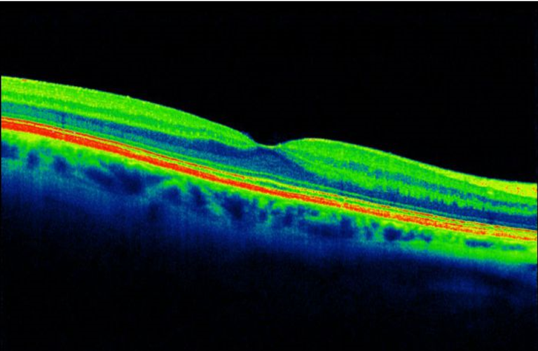
OCT uses light waves to take cross-section pictures of retina’s distinctive layers. This helps to map/ measure the thickness of retinal layers and in turn helps diagnose the severity and nature of damage in retina.
Scan report from OCT
Fundus Imaging
Fundus imaging involves photographing the rear of an eye; also known as the fundus. The main structures that can be visualized on a fundus photo are the central and peripheral retina, optic disc and macula. Fundus imaging is used to inspect anomalies associated to diseases that affect the eye, and to monitor their progression. It is able to identify glaucoma, as well as monitor disease processes such as macular degeneration and diabetic retinopathy.
Naveen Eye Hospital is equipped with OCT scan and Fundus imaging facilities to help with correct diagnosis and comprehensive treatment.